
How to represent 3D space in the entorhinal cortex of flying bats?
Authors
*G. GINOSAR1, J. ALJADEFF2, Y. BURAK3, H. SOMPOLINSKY3, L. LAS1, N. ULANOVSKY1;
1 Weizmann Inst. of Sci., Rehovot, Israel; 2 Dept. of Bioengineering, Imperial Col. London, London, United Kingdom; 3 The Edmond and Lily Safra Ctr. for Brain Sciences, and Racah Inst. of Physics, Jerusalem, Israel
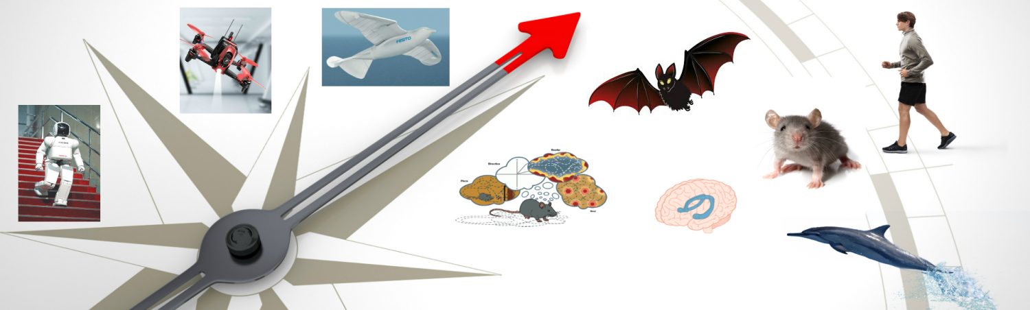
“Grid cells are neurons in medial entorhinal cortex (MEC) that are activated when the animal passes through multiple locations (‘firing-fields’) on the 2D surface that the animal is exploring. These firing-fields are arranged in a hexagonal 2D lattice that spans the entire 2D surface. Although many animals navigate in 3D space, the volumetric 3D firing-pattern of grid cells remains unknown. Here we recorded MEC neurons in freely-flying bats, and found a variety of spatial cells – including 3D border-cells, 3D head-direction cells, and neurons with multiple 3D firing-fields. The multi-field neurons displayed an increased inter-field-spacing along the dorso-ventral axis of MEC – as in rodent grid-cells. Many of the multi-field neurons were 3D grid cells – exhibiting a local order in their field arrangement, with fields separated by a characteristic distance; however, they were not organized in a global lattice. We modeled grid-cells as emerging from pairwise-interactions between fields, which yielded a hexagonal lattice in 2D, but only local order in 3D – thus explaining both 2D and 3D grid-cells within one model.“
For further info, please visit https://www.abstractsonline.com/pp8/#!/7883/presentation/64763
About
CogNav Blog
New discovery worth spreading on cognitive navigation in neurorobotics and neuroscience
Recent Posts
- How to build a bio-inspired hardware implementation of an analog spike-based hippocampus memory model?
- How does the brain select what to remember during sleep?
- How hippocampal activity encodes numerous memories of specific events in life?
- How egocentric coding properties arise from its presynaptic inputs, and how egocentric cells represent items across different behavioral contexts?
- How the medial entorhinal cortex develops during learning and influences memory?
Tags
Categories
- 3D Movement
- 3D Navigation
- 3D Path Integration
- 3D Perception
- 3D SLAM
- 3D Spatial Representation
- AI Navigation
- Bio-Inspired Robotics
- Brain-Inspired Navigation
- Cognitive Map
- Cognitive Navigation
- Episodic Memory
- Excerpt Notes
- Flying Vehicle Navigation
- Goal Representation
- Insect Navigation
- Learning to Navigate
- Neural Basis of Navigation
- Path Integration
- Path Planning
- Project
- Research Tips
- Robotic Vision
- Self-Flying Vehicles
- Spatial Cognition
- Spatial Cognitive Computing
- Spatial Coordinate System
- Spatial Memory
- Time
- Unclassified
- Visual Cortex
- Visual Cue Cells
Links
- Laboratory of Nachum Ulanovsky
- Jeffery Lab
- BatLab
- The NeuroBat Lab
- Taube Lab
- Laurens Group
- Romani Lab
- Moser Group
- O’Keefe Group
- DoellerLab
- MilfordRobotics Group
- The Space and Memory group
- Angelaki Lab
- Spatial Cognition Lab
- McNaughton Lab
- Conradt Group
- The Fiete Lab
- The Cacucci Lab
- The Burak Lab
- Knierim Lab
- Clark Spatial Navigation & Memory Lab
- Computational Memory Lab
- The Dombeck Lab
- Zugaro Lab
- Insect Robotics Group
- The Nagel Lab
- Basu Lab
- Spatial Perception and Memory lab
- The Neuroecology lab
- The Nagel Lab
- Neural Modeling and Interface Lab
- Memory and Navigation Circuits Group
- Neural Circuits and Memory Lab
- The lab of Arseny Finkelstein
- The Epstein Lab
- Gu Lab (Spatial Navigation and Memory)
- Fisher Lab (Neural Circuits for Navigation)
- The Alexander Lab (Spatial Cognition and Memory)
- Harvey Lab (Neural Circuits for Navigation)
- Buzsáki Lab
- ……